Category: Cancer

Choosing the RIGHT Metrics for Dashboards

Dashboards are effective tools to communicate data, but they can also be misleading or confusing if the wrong metrics are used. So, how do you choose the right metrics or indicators?

Here is a short article with 6 steps to help you get started. Click HERE.

What is Cardio-Oncology? (Video)

Cancer and cardiovascular disease are the two most common causes of death. The association between cardiotoxicity and cancer treatment has been known for some time. But, cardio-oncology as a medical subspecialty is fairly new. Cancer Registrars should understand the risks, short- and long-term side effects if they are to accurately review and document the patient’s continuum of care. To get you started here is a short 3.5 minute video.

T-Cell Therapy – Cancer Breakthrough? (Video)

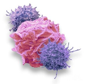
Can cancer patients skip conventional chemotherapy and radiation therapy treatments and undergo T-cell therapy instead? New research conducted by UCLA, Stanford and others shows promise. Watch this video below.

Skip The Chemo: Cancer Breakthrough | Watch (msn.com)

Strategies to Lower Cancer Risk

One of the biggest challenges faced with conducting cancer prevention events is establishing a knowledge base and understanding with the participants. To create a healthier population, or to educate the community in such a way that they take a proactive step to living a healthier life, it is important to develop common-sense strategies that the community understands. It’s important to teach them about sun exposure and skin cancer risk, but equally important to teach them about other risk reduction strategies, such as exercise for busy moms or simple, cost effective food preparation.

There are no guidelines or limitations to the type of strategies you can develop. What is important is that you understand your communities unique needs and lifestyle norms so you can tailor the strategy to what is happening right where you live. Some research notes that lack of “real-world” resources or education that is focused on the population are barriers to delivering an effective program.

In this article methods to create population-specific strategies are discussed. Share this with your cancer committee, community outreach coordinators or other individuals who will help you plan your events.

To read the Oncology Nursing Society (ONS) article click here.

Registries & Cervical Screening

Cervical cancer screening helps to detect early disease and saves lives! And yet, according to the CDC, over 13,000 women are diagnosed each year and over 4,000 will die from their disease. About half the women who are diagnosed with cervical cancer never received screening.

The CDC launched a study to look into cervical cancer screening. Cancer Registries in Michigan, New Jersey and Louisiana participated in the study to identify women who were 21 years of age or older and diagnosed with cervical cancer. Patients were surveyed about whether they were screened or not, and if not, the reasons why. Household income, health insurance, race and ethnicity were also collected.

The study determined that of the women who participated, over 60% of them had not undergone screening for early detection. At the top of the list of reasons were that the women were not aware of the risk factors, understand test procedures or results, or did not have insurance benefits to pay for testing.

The study analysis included a lot of other valuable information as well that can be used by cancer centers and programs to develop cervical cancer screening and community outreach programs. To read the entire article on the CDC website, click here.

Lymphovascular Invasion – Synonymous Terms

There is no shortage on synonymous terminology in the medical record. Knowing whether to accession a case, or how to code the extent of disease, in a case that is laced with synonymous or ambiguous terms can be daunting to say the least! Fortunately, there are standard-setting agencies guidelines to help the Cancer Registrar make the right determination and assign proper codes to the case abstract.

Recently a query was posted to the Surveillance, Epidemiology and End Results (SEER) SINQ inquiry system (SINQ #20210036) that caught our eye. Paraphrased, the question was: are lymphovascular invasion and lymphovascular space invasion the same term or do they describe something different? Great question!

So we went to our favorite pathology dictionary and looked it up. There we learned that lymphovascular invasion, or LVI, is the term given to the movement of cancer cells into either a blood or lymphatic vessel. Once the cancer cells make their move they have the ability to spread to other parts of the body. Cancer Registrars know this movement to another part of the body to be called metastasis.

Lymphovascular invasion is an important prognostic indicator that pathologists may see in the tissue sample. Pathology reports stating the specimen is positive for lymphovascular invasion mean that it was seen in the tissue examined and conversely, if stated as being negative it means it was not seen.

You will be happy to know that according to SEER, the two terms, lymphovascular invasion or lymphovascular space invasion are synonmous with one another.

Now, just in case you’re interested in what the pathologist sees when he/she finds lymphovascular invasion, we found an example from specimen slide on the UCDavis Department of Pathology website. In the graphic below you can clearly see the malignant cells (small dark circles or dots) spilling out of the lymphatic space (white) and into the surrounding tissue.

OLYMPUS DIGITAL CAMERA

So now you know the difference between the two terms!

Cancer A to Z

There are over 300 different types of cancer. A cancer registrar is specially trained in how to correctly gather the data from the medical record and to code and classify it for research and strategic planning. From time to time they will come across a type of cancer they may not be as familiar with as compared to cancers like lung, colon, prostate, breast, etc. Having a resource like this one published by the National Cancer Institute (NCI) is very helpful.

What Are Cancer Statistics?

Cancer Registrars collect and analyze data to tell a story about the latest trends in their facility, region, state or nationally. Understanding how the statistics are derived and calculated, especially if you are going to compare your hospital with another program, state or national benchmark is very important.

SEER (Surveillance, Epidemiology and End Results) has made available a series of online videos called Did You Know? The video’s highlight key topics and trends and are helpful in understanding cancer-related statistics.

Here is one of these videos, titled “Cancer Statistics.”

Cancer Statistics | Did You Know? | Surveillance, Epidemiology and End Results (SEER)

When is CLL in Remission?

When is chronic lymphocytic leukemia (CLL) in remission? What does it mean when the medical record states the CLL patient has a complete or partial remission? All great questions often heard around the table when cancer registrars get together.

CLL is the most common type of leukemia in the United States in American adults. Over 175,000 men and women are currently living with CLL, and many of them with a good quality of life. While a cure is not yet available there are a wide-range of effective treatments available today. For patients with slow-growing disease or who are in clinical remission, they may not even require active therapy. Of course, Cancer Registrars will be looking for all of this information and coding it appropriately in the case abstract.

Standard treatment for CLL is chemotherapy and radiation. However, newer treatments are being tested and made available to patients to help them achieve partial and complete remission for longer and longer periods of time. Newer therapies include:

  • Immunotherapy:
    • Drugs that work with the body’s immune system to attack the cancer cells similar to how it attacks other diseases.
  • Chemotherapy:
    • Treatment regiments combined with stem cell transplant are being used to kill the cancerous cells in the body and are then replaced with healthy cells via a stem cell transplant.
  • Targeted Drugs:
    • These new medications attack specific substances that help the cancer cells to grow and multiply and leave the healthy, non-malignant cells alone.

There are two types CLL remission which we will describe below:

  • Complete:
    • If the patient’s blood tests no longer show the presence of CLL and they do not have any symptoms, such as swelling in the lymph nodes or spleen, then CLL is considered to be in complete remission.
  • Partial:
    • If the patient is symptom-free (i.e., no swelling in lymph nodes) but there is still a small amount of CLL detected in their blood tests, then they are considered to be in partial remission.

Laboratory tests are an important part of the medical record that the Cancer Registrar must review. For CLL you should look at the following:

  • Blood Testing
    • Diagnosing CLL usually starts with a routine blood test called complete blood count, or CBC. A CBC measures a number of different types of cells in the sample. For example, a patient may have CLL if the blood contains too many white blood cells, or WBCs. The CBC also measures red blood cells (RBCs) and platelets. Low levels of RBCs is called anemia and low platelet count is called thrombocytopenia.
  • Flow Cytometry and Cytochemistry
    • For these tests chemicals or dyes may be applied to the blood sample in the laboratory to provide information about the type and subtype of leukemia. CLL has distinctive markers, called cell surface proteins, on the outside of the cell. The pattern of these markers is called immunophenotype and is used to distinguish CLL from other types of leukemia that also involve lymphocytes. Flow cytometry, also called immunophenotyping, is the most important test used to confirm a diagnosis of CLL.
  • Genomic and Molecular Testing
    • These tests look for specific genes, proteins, chromosome changes or other factors unique to leukemia. Because CLL cells divide very slowly, looking at chromosomes is often less useful than tests that find specific genetic mutations or changes. Fluorescence in situ hybridization (or FISH) assays and other genetic tests, such as polymerase chain reaction, are used to find genetic mutations. Some of the genetic changes that are found in CLL include (but are not limited to):
      • Deletion of the long arm of chromosome 13 [del(13q)], which is found in about half of all CLL patients,
      • An extra copy of chromosome 12 (trisomy 12),
      • del(11q),
      • del(17p),
      • NOTCH1 mutations,
      • SF3B1 mutations,
      • TP53 abnormalities,
      • MYD88 mutations, and
      • IGVH, which may be important whether it is changed or unchanged.
    • Genomic and molecular testing can be used to determine how quickly the disease will progress and to identify treatment therapies and options. For example, if a patient has del(17p) genetic mutation then they are more likely to have a leukemia that is difficult to treat and some may work better than others.
  • Imaging
    • It is common for CLL to be found in many different parts of the body, even if diagnosed early. Imaging tests are rarely used to diagnose CLL but they may be used before treatment is given to identify areas of involvement or to determine how well CLL is responding to treatment.
  • Bone Marrow Aspiration and Biopsy
    • Bone marrow biopsy is also not generally used to diagnose CLL, but it may be done before treatment starts. It is useful to help determine the prognosis or a patient’s chance of recovery.

CLL can be in remission for many years, but there is always a chance it could come back. It is not uncommon for a physician to recommend a “watch and wait” form of long-term surveillance. This is why it is so important for the Cancer Registrar to understand the disease process and to gather accurate and complete follow-up and cancer status information throughout the patient’s lifetime.

Cancer Registrar’s should consult the SEER hematologic coding and abstracting resources to determine other steps or actions taken during the case abstraction process, whether they are in a SEER-designated State or not.

Disclaimer – this article is not used to determine coding or abstracting standrds! Rather it is an informational guide to help the Registrar understand what they may find in the medical record. For coding or abstracting standards please refer to your apppropriate manuals.

What Are Biomarkers?

Biomarkers, or biological markers, are molecules found in body tissues and fluids, including tissue and blood. Cancer biomarkers provide important information about the patient’s tumor. Biomarker terminology varies but it may also be called biomarker testing or tumor testing. Biomarkers are usually a protein or antibody that is released by the tumor, or they may be the body’s response to the presence of cancer, which shows up like a gene mutation. There are a number of biomarkers used by clinicians today and more are being studied.

When a clinician orders a biomarker study, they are looking to answer important questions about the patient’s disease that may include:

  • How aggressive is this cancer?
    • Prognostic biomarkers tell us about the estimated course, or progression, the cancer will take if it is not treated. These markers help to identify how well organized the cell is, or if it is not functioning at all.
  • What is the best drug to use to treat this cancer?
    • Predictive biomarkers are ordered to identify the best available treatment because they may preduct whether or not the cancer will respond to a specific treatment.
  • Will this cancer recur, or when will it come back?
    • Recurrence biomarkers may be used to monitor the presence of a cancer or whether it has recurred after initially responding to treatment.
  • What type of cancer does the patient have?
    • Diagnostic biomarkers aid in identifying the type of cancer a specific patient may have.
  • What is the correct dose of drugs needed to treat an individual patient?
    • Pharmacodynamic biomarkers are useful to determine the dose, usually chemotherapy, that will be given to the patient.

As you can see above, biomarker testing helps the clinician identify critical information needed to develop and deliver an effective plan of treatment. Testing should happen as close to the diagnosis as possible and before a treatment plan is launched.

Discussion of biomarker results or as a predictor for treatment or recurrence is included in multidisciplinary cancer conferences (aka tumor boards) and physician-to-patient discussions. Cancer Registrars gather biomarkers and include relevant values and their types in the cancer case abstract that is used for clinical research, quality studies and comparative analysis.

(Source: Biomarkers and Biomarker Testing, Fight Colorectal Cancer, Springfield, MO.)


Warning: Undefined array key "student_url_profile" in /home3/mwebb/public_html/wp-content/plugins/masterstudy-lms-learning-management-system/_core/lms/helpers.php on line 1340

Warning: Undefined array key "student_url_profile" in /home3/mwebb/public_html/wp-content/plugins/masterstudy-lms-learning-management-system/_core/lms/helpers.php on line 1345